

Lasers for Cultural Heritage
2018 is the European Year of Cultural Heritage. A good time to highlight some of the fascinating cultural heritage projects executed with the help of laser experts from Laserlab-Europe. As this focus section shows, lasers can be of unique value for the analysis of objects that are part of our common European cultural heritage – ranging from coins dating from the Roman Empire to gemstones from the Late Medieval Period and illustrious painters like Peter Paul Rubens. Our Greek partner IESL-FORTH, represented in this focus with two different contributions, is a very active partner in the EU-funded IPERION CH project (formerly CHARISMA), which aims at establishing a European infrastructure for restoration and conservation of cultural heritage. This involvement has led to the creation of a Mobile Access Facility specifically tailored for analysis of cultural heritage objects. It is worth noting that also Laserlab-Europe subcontractor INO-CNR (Italy) and associate partner WIGNER-RCP (Hungary) are involved in IPERION-CH.
SORS spin-off as a tool for investigation of heterogeneous painted systems (CLF, UK)
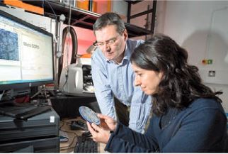 |
| Prof. Pavel Matousek and (CLF) and Dr. Claudia Conti (CNR) ©STFC |
Micro-Spatially Offset Raman Spectroscopy (micro-SORS), a recently developed technique, has been used by teams from Italy and the UK’s Central Laser Facility (CLF) for the non-destructive chemical characterisation of stratified paint samples. The results, published in the journal Analytical Chemistry, provide new opportuni- ties for cultural heritage research, often where heterogeneous layers are found within painted stratigraphies.
Since its invention by Indian physicist C.V. Raman in 1928, a new technique based on the Raman concept – but with significantly higher penetration depth – has emerged. If you combine SORS with the power of microscopes, you get a technique known as Micro-Spatially Offset Raman Spectros- copy, or micro-SORS for short, developed by teams of the Italian National Research Council (ICVBC-CNR) and CLF.
As it has very high chemical specificity, micro-SORS is often used for the characterisation of surface layers of paints in art. Not only that, but the technique has come in very handy for the study of hidden images and writings, as it can determine overlayer depth, reject overlayer fluorescence and permit two-dimensional mapping of thin materials.
In this experiment, scientists from the CNR and CLF studied two-layer paint systems where either one or both layers were heterogeneous, that is, they were made up of more than one type of material, in this case, paint.
Using micro-SORS technology, the team were able to obtain informative data on the overall chemical makeup of the sample. They then tested the behaviour of micro-SORS signals under different scenarios. In order to do so, the team plotted the ratio of the top layer signal over the sublayer sig- nal as a function of defocusing distance for each of the two subsurface components from data obtained by mapping on scales substantially larger than the scale of heterogeneity. This approach provided an effective means of obtaining robust and representative micro-SORS datasets from which sample composition could be effectively deduced, even in the extreme scenarios of high heterogeneity.
The analysis of paint samples is not the only application of this research, however. In fact, this technique is very applicable to a wide range of areas including – but not limited to – polymer research, forensics and biological fields. Whilst the paper does raise concerns about the effectiveness of micro-SORS with highly heterogeneous samples, informative data on the overall chemical makeup of the sample in the team‘s experiment was successfully obtained. This means that micro-SORS is quickly becoming a valuable tool for non-invasively investigating the chemical composition of subsurfaces and thus providing detailed insight into turbid layers such as those found in paintings and other man-made historical artefacts.
Emily Cooke
C. Conti et al., Analytical Chemistry 89: 11476-11483, 2017
Raman spectroscopy applied to the Treasure of the Queen Saint Isabel (Coimbra Laser Lab, Portugal)
 |
|
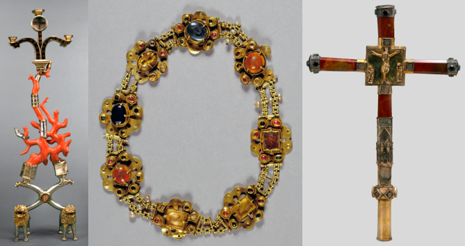
Image of the Treasure of the Queen Saint Isabel (from left to right): The Reliquary of the Holy Cross, the Necklace
and the Processional Cross. ©Machado de Castro National Museum
|
Known as the Treasure of the Queen Saint Isabel, the 14 th century bequest of Isabel of Aragon, Queen of Portugal, is one of the most valuable sets of art pieces in Portuguese history. The Treasure is part of the Machado de Castro National Museum collection, and had not been studied extensively by analytical characterisation techniques to this date. Recently, the Treasure has been analysed by a team from Coimbra Laser Lab, in order to identify the gemstones, pigments and the presence of vitreous enamel.
The study of these pieces was performed by micro- Raman spectroscopy, with laser excitation of 532 nm and 633 nm. This technique couples Raman spectroscopy with optical microscopy, allowing us to focus on the samples’ areas of interest without sample preparation and in a non- invasive and non-destructive way. The sensitivity and selectivity of the technique were also of prime importance to the performed investigation. The vibrational signatures characteristic of each of the studied materials were obtained and compared with Raman spectra databases, providing a reliable, fast way to characterise the different components of the artworks investigat ed. Detailed results have been described as part of a Thesis.
The Necklace (in the centre of the image) is composed of eight multi-lobed plates, garnished with gems and linked by a chain, which are decorated with baroque pearls. According to the legend, this is the surviving part of a necklace of St. Isabel that, because it was considered to be able to provide miraculous gifts, was damaged and degraded by the sick people – especially women in labour – anxious for a fragment as a holy relic that would protect them. All of the stones and the pearls were successfully identified, providing an extensive characterisation of this piece of jewellery regarding this characteristic.
The Processional Cross (right image) combines blood-red jasper with gilt silver and the blue of the processional stones, an excellent frame for the Christian theme represented on the central crossing. The gems and the jasper of the cross were analysed, as well as the presence of vitreous enamel in specific parts of this artwork, providing a better understanding of its possible origin within the Iberian Peninsula.
The Reliquary of the Holy Cross (left image), made from silver and coral, decorated with coloured enamels and imprinted with the arms of the kingdoms of Portugal and Aragon, is a rare piece without parallel regarding its form, and laden with symbolism and evocations. The enamels that decorate the upper section of the Reliquary of the Holy Cross are translucent (a technique used in the workshops of Avignon and Montpellier), and were identified, as well as the coral, which is one of the major components of the piece.
Bernardo A. Nogueira, Ana Peneda, Rui Fausto (Coimbra Laser Lab), Fernanda Alves, Pedro Ferrão (Machado de Castro National Museum – Coimbra)
A. Peneda, Autenticação de Obras de Arte por Microscopia de Raman, M.Sc. Thesis, U. of Coimbra, Portugal, 2017
Analysis of semiconductor pigments by photoluminescence microscopy(CUSBO, Italy)
 |
|
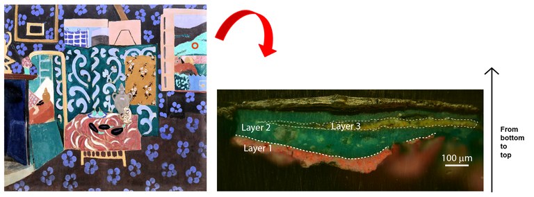
Figure 1: (Left panel) Colour image of the painting Still life with eggplant by Henri Matisse (Musée des Beaux-Arts, Grenoble, France,
210 x 245 cm (1911)). The location of microsampling is indicated by the red open circle. (Right panel) Colour image of the stratigraphic
microsample composed of a bottom orange layer (layer 1), a thick green layer (layer 2) and a yellow layer (layer 3). The microsample
is shown in its upright position (as stated by the black arrow) with the inner layers of the painting being displayed at the bottom of the image.
© Comelli et al. 2017 |
Many works of art, such as paintings by Van Gogh, Picasso, Monet, and Cézanne, contain semiconductor pigments. At Laserlab-Europe partner CUSBO, a time-resolved photoluminescence (TRPL) microscope was recently developed for the analysis of cross-sections of paint layers made of luminescent semiconductor pigments.
Illustrative examples of semiconductor pigments include ancient pigments, such as the brilliant red vermillion, first used during the Neolithic Age, and the lemon yellow orpiment, extensively found on ancient Egyptian objects and paintings. Thereafter, a variety of new semiconductor pigments were produced in the Modern Age – starting from the second half of the 19 th century – when synthetic semicon- ductor pigments were introduced, and include zinc white, zinc sulphide, titanium white and cadmium yellow and red.
These synthetic pigments were extensively used by painters as Van Gogh, Picasso, Monet and Cézanne, since they offered new hues and greater covering power than many paints based on natural minerals. However, these pigments were often used with few concerns about their chemical instability, which was often the cause of paint degradation and discolouration. Indeed, recent research has highlighted the complexity and the chemical instability of some historical semiconductor pigments, elucidating the presence of trace metal impurities and of various reaction products. In this context, photoluminescence is a unique method for the detection of luminescent trace impurities and compounds, heterogeneously present on a painted surface and in historical samples, whose presence can be linked to the methods used for pig- ment synthesis or to paint degradation.
At the CUSBO facility, we have recently d eveloped a time-resolved photolumines- cence (TRPL) microscope, equipped with both spectral and lifetime sensitivity at timescales ranging from nano - seconds to hundreds of microseconds, for the analysis of cross-sections of paint layers made of luminescent semi- conductor pigments.
Despite being widespread amongst conservators and museum curators, the analysis of the fluorescence emission from samples and artworks is usually employed as a visual and qualitative method for documenting the presence of luminescent compounds. Similar considerations can be reported for the use of fluorescence optical microscopy, which is occasionally applied to stratigraphic samples of paintings simply for putting light on sample heterogeneities.
In contrast, here we illustrate the potential of TRPL microscopy for the characterisation of a micro-sample from the painting Still Life with Eggplant (Musée des Beaux-Arts, Grenoble, France) by Henri Matisse (1869-1954) (Figure 1 – left panel). The TRPL method, taking advantage of spectral and lifetime-resolved data, has provided us with valuable information for the identification of semiconductor pig- ments in paints. In particular, in the present case study we demonstrate the identification of the semiconductor pig- ments zinc white and cadmium yellow in different layers of the microsample.
Daniela Comelli
D. Comelli et al., Materials 10: 1335, 2017
Portable holographic interferometry system for detecting surface deformation (IESL-FORTH, Greece)
 |
|
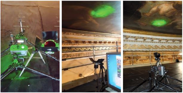
The Laser interferometry DHSPI system setup facing the ceiling during examination of paintings by Peter
Paul Rubens at the Whitehall Banqueting House in London (Photos: IESL-FORTH). |
Digital Holographic Speckle Pattern Interferometry (DHSPI) is a non-destructive optical technique which can be used to investigate deformation, deterioration and fracture mechanisms in cultural heritage objects. In the context of the European IPERION CH MOLAB project, Laserlab-Europe partner IESL-FORTH has developed DHSPI-II, a portable custom-made prototype, which has already been used to monitor wooden objects and to examine 17 th century paintings by Peter Paul Rubens at the Whitehall Banqueting House in London.
In DHSPI two coherent beams from the same laser form a holographic interferogram of the speckle patterns – spatial variations in the observed light intensity of the light reflected from a rough surface – on a CCD detector. A computer processes the phase changes in the reflected beam, which are caused by changes in the shape and position of the surface and the underlying material, revealing any changes in the surface as well as the internal structure.
The laser beams employed in the DHSPI-II are highly divergent, allowing imaging of the whole or extended parts of the surface (full field). This also guarantees safety for the operator as well as the object, because in this power density regime the laser radiation does not interact with the surface. The technique enables detection of the location, shape and size of invisible defects, as well as their changes over time. As such, it allows monitoring of structural changes resulting from varying environmental and climatic conditions, conservation treatments, aging, and transportation or handling. The DHSPI-II instrument is a 15 kg light-weight portable system, comprising an optical head of 30x20 cm and a PC driven control unit. It has been developed at the holography laboratory of IESL-FORTH, and funded through several national and EU projects related to artworks diagnostics.
In a preliminary preventive conservation experiment, DHSPI was employed to monitor in real time the response of complex materials and objects to fluctuations in relative humidity and temperature. Analysis has focused on the assessment of the dynamics of dimensional changes in cultural heritage objects made of hygroscopic materials. Oak and pine with sizes of 2 x 2 cm, and of varying thicknesses from 1 mm to 1 cm, were monitored while relative humidity was varied between 45% and 65% at different rates. The data of this work in progress suggests that wood sensors could be suitable for future assessment of environmental changes in collections.
In addition, scaffold access between February and April 2018 enabled the recording of The Apotheosis of King James I and The Wise Rule of King James I, two ceiling paintings produced by Peter Paul Rubens and installed at the Whitehall Banqueting House in London in 1636. The assessment by the DHSPI-II instrument was part of a multidisciplinary effort to carry out a first ever full and systematic technical conservation survey, to determine how the paintings were created, how they have changed over the years, as well as to establish an accurate record of the present condition of the paintings to inform on approaches to future possible conservation interventions.
Vivi Tornari
A. Nevin, M. Andrianakis, V. Tornari, Towards understanding the impact of environmental changes on cultural heritage through the real time monitoring of dimensional change in wood sensors with Digital Holographic Speckle Pattern Interferometry, to be published in Applied Surface Science
Listening to laser light interactions with objects of art: a photoacoustic approach (IESL-FORTH, Greece)
 |
|
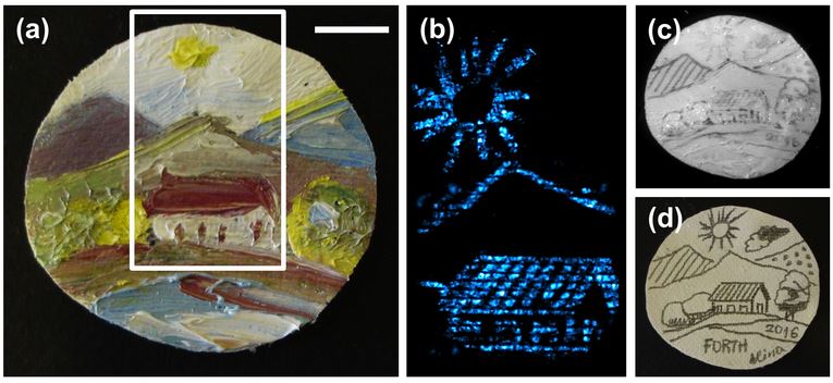
Photoacoustic detection of underdrawings in miniaturepaintings. (a) Brightfield view of a miniature painting (rurallandscape).
(b) Photoacoustic image of the painting’s underlyingsketch over a central region of 2.2× 3.8cm as indicated by thewhite box in (a). (c) NIR image of the underdrawing at 1200nm. (d) Brightfield view of the original pencil sketch prior to overpainting. Scalebar: 1 cm. (Adapted from Tserevelakis et al. 2017) |
Photoacoustic imaging is a novel, rapidly expanding diagnostic technique, which has been predominantly developed in the context of contemporary biomedical research. At IESL-FORTH, it has recently been demonstrated that photoacoustic imaging can break the barriers of biomedicine and find innovative applications in diagnostics of cultural heritage objects.
In photoacoustic imaging, pulses of laser light are typically used to locally heat a material, inducing ultrasonic sound waves as a result of rapid thermoelastic expansion. Because the magnitude of these acoustic vibrations depends on the local absorption properties of the material for the selected irradiation wavelength, the produced waves can be used to derive an optical absorption map of the examined piece of material. Using ultrasonic transducers as detectors, images can be constructed of the area exposed to the laser light.
Because the photoacoustic signal has over three orders of magnitude higher transmission through strongly scattering media compared to light in the visible and near infrared, it offers substantially better detection sensitivity and achieves excellent optical absorption contrast at high spatial resolution. Such an inherently hybrid approach, combining light with ultrasound, allows for new possibilities in the diagnosis of artworks, by overcoming several limitations of purely optical techniques mostly related to light scattering.
The unique capabilities of photoacoustic imaging have already been exploited to establish a radically new non-destructive methodology for the uncovering and differentiation of well-hidden features in multi-layered cultural heritage objects such as paintings and documents. It can, for instance, be used to image underdrawings of paintings, which are of great interest to art historians and cultural heritage scientists alike.
Furthermore, it has been demonstrated that the attenuation of generated photoacoustic signals during their propagation through optically opaque media (e.g. paints) can determine precisely the thickness of thin layers, providing micrometric precision stratigraphic information on the artwork under investigation. Finally, photoacoustic signal detection has been ex - ploited for the in situ real-time monitoring of laser cleaning interventions, promoting an improved conservation outcome by safeguarding artworks’ original surfaces.
George Tserevelakis
G.J. Tserevelakis et al., Scientific Reports 7: 747, 2017
Analysis of bronze coins by LIBS (Institute of Physics, Croatia)
 |
|
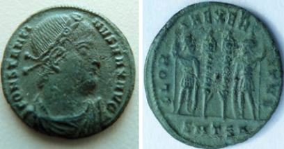
A coin made for Constantinus I the Great in the Thessalonica
mint analysed by LIBS (Taken from Orlić Bachler et al. 2016) |
Ancient coins can provide valuable information about the technology and economy of the society in which the coins were minted and used. In a recent study, a team from Laserlab-Europe associate partner Institute of Physics (Zagreb, Croatia) has applied Laser-Induced Breakdown Spectroscopy (LIBS) for experimental investigation of the elemental composition of, and correlations between, antique bronze coins coated in silver – known as follies – dated from the Roman Empire. The coins were made in different mints during the reign of different rulers.
Numerous non-invasive or almost non-destructive experimental techniques are available to obtain qualitative and/or quantitative information about unknown materials. LIBS is especially valuable for the analysis of ancient coins, as it can be used to retrieve the elemental composition inside the bulk of the coin, which has not been affected by corrosion or influenced by modern conservation methods. This work presents a feasibility study of applying Principal Component Analysis (PCA) to data obtained by LIBS with the aim of determining the correlation between different samples.
Every LIBS spectrum contains a variety of complex information exploitable for qualitative and quantitative analysis of the elemental content of the sample. For an efficient analysis of complex LIBS spectra, as well as for the assessment of the analytical capabilities of this experimental method, it is necessary to use appropriate statistical methods.
In this study, experimental LIBS data were analysed by multivariate analysis. Principal Component Analysis (PCA) showed that for each sample, the ablation spectra can be grouped together based on their prominent spectral features. This grouping is connected to the progression of ab - lation from the surface to the bulk of the sample. Also, by using PCA the samples were grouped according to similari- ties/differences in elemental composition. This procedure revealed which elements, as far as the emission spectrum is concerned, make samples (dis)similar. The method of Partial Least Squares (PLS) was used to estimate the quantitative com- position of the samples. By observing changes in the ratio of intensities of silver and copper lines we determined similari- ties and differences in the material that was used in the mint Siscia during the reign of vari- ous rulers. In the period 286 to 383 CE, the portion of silver in follies was generally decreasing as expected. Using the PLS method we found that copper concentration in the analysed ancient coins ranged from 83% to 90%.
Our investigation shows that LIBS, performed under argon flow to suppress the air signature in the spectrum, is a suitable method for analysis in the field of numismatics in particular and cultural heritage in general, while application of the multivariate analysis method is important for more complete analysis and better understanding of complex data. Moreover, damage caused by ablation of the surface was limited to micrometres and was therefore considered insignificant, making LIBS a non-destructive technique for analysis of delicate samples.
Niksa Krstulovic
M. Orlić Bachler et al., Spectrochimica Acta Part B 123: 163-170, 2016
Laser rejuvenation of ancient Greek masterpieces (IESL-FORTH, Greece)
 Laser cleaning of the Caryatids is taking place in situ at their exhibition site in the Acropolis Museum © The Acropolis Museum, Athens, Greece |
Our Greek partner IESL-FORTH has developed a laser system capable of cleaning ancient sculptures. In January 2011, the cleaning laser system was set up on the visitors' floor of the Acropolis Museum, where the Caryatids – sculpted female figures originally serving as columns or pillars – are displayed to the public. Since museum policy is to avoid risky transportation of the precious pieces from ancient Greece, removal of pollution accumulation now takes place under the eyes of the visitors - naturally behind a protective housing. The laser cleaning technique was selected from over forty competing methods, and has already been successfully used to clean other Athens Acropolis Sculptures - such as the Parthenon West Frieze and the Frieze of Athena Nike's temple. The laser system is operated at infrared and ultraviolet wavelength simultaneously and can be used to remove thick layers of pollution in a controlled and safe way. The combination of the two wavelengths ensures that the surface of the pieces of art treated is not damaged and the colours are not affected. This unique laser system was developed in close collaboration with the Acropolis Restoration Service and the Ephorate of Prehistoric and Classical Antiquities in Athens. IESL-FORTH has a long history of developing innovative laser and optical technologies for conservation and diagnostics of works of art. Researchers of IESL-FORTH have developed mobile laser systems that are used to map layers of paint and visualize underdrawings, and laser spectroscopy is employed to identify pigments in paintings, icons and illuminated manuscripts, as well as for analysis of archaeological metal, glass and pottery objects. In a holographic approach, IESL-FORTH researchers use expanded laser beams to uncover effects of deterioration within the structure of objects of art. With this holographic technique, defects that are not visible by other means, including X-ray imaging, become quantifiable. |
UV-fluorescence spectroscopy for identification of varnishes in works of art (CUSBO, Milano)
 |
|---|
|
Fig. 1: The portable FLIM instrument developed at the Politecnico |
Darkening and yellowing of a varnish film due to aging is one of the main conservation problems in paintings and the varnish film may need to be periodically removed and replaced with a new one. Hence, the characterization of the varnish layer is essential in the planning of a restoration work, in order to choose the most suitable solvent or cleaning method for its removal. In this field, both the Italian research group at Physics Department of Politecnico di Milano, headed by prof. Rinaldo Cubeddu, and the French research group from the Institut des NanoSciences de Paris, headed by prof. Mady Elias, have extensive expertise in the analysis of works of art with optical spectroscopy techniques. Thanks to the LASERLAB-EUROPE facilities, the two research groups have been able to initiate a fruitful collaboration by employing their complementary fluorescence spectroscopy devices for the non-destructive analysis of a varnished painting.
Transparent varnished coatings have long been used on paintings for both protective and aesthetic purposes: a varnish layer, in fact, protects a paint film from dirt and mechanical damage, while at the same time saturating colours because of the refraction index matching.
A varnish layer is basically a film, of only microns in thickness, often made of a natural or a synthetic resin diluted in a suitable solvent. As an example, triterpenoid resins, such as mastic and dammar, used on their own or mixed with wax and oils, were the most popular varnishes because of their excellent adhesive properties, their good solubility in solvents, and because they yellow to a lesser extent than varnishes made with other resins. In most cases, the determination of the chemical composition of a varnish layer is carried out in laboratory using sensitive and highly specific chromatographic-mass spectroscopy techniques. However, the costs and extensive work-up associated with these analyses are significant; furthermore, sampling of varnish layers is not always straightforward. Therefore, alternative and complementary non-destructive techniques are particularly attractive.
With the advantage of not requiring any sample and being non-contact, UV-induced fluorescence is a useful phenomenon which can be used for the non-invasive examination of works of art, since many artists' materials, including binding media, modern pigments and colorants may be luminescent. Due to their chemical composition, even varnishes are naturally fluorescent materials or can develop fluorescence upon ageing. Hence, the visual examination of the fluorescence emitted from a varnished painting upon excitation with a simple UV lamp is widespread, providing immediate indications to conservators and curators regarding the presence of retouching, repainting and surface heterogeneity. Beside this simple approach, in recent years diffe- rent laboratories have developed spectroscopic portable devices for the in situ analysis of works of art with features almost comparable to those of standard laboratory equipments.
The instrument developed in the French CNRS laboratories is a portable UV and visible spectrometer, capable of measuring both the fluorescence and the diffuse reflectance spectrum of a point of interest on a painted surface. A database of fluorescence spectra of known aged varnishes have been built up for varnish identification. Up to now, the database contains over 110 fluorescence spectra of diffe- rent aged resins and varnishes, representative of the varnish layers most commonly found on easel paintings. The fluorescence spectrum of the unknown varnish can be numerically compared with the spectral database using a least square method that prioritizes the wavelength range of the maxima of the reference spectra.
Researchers at Politecnico di Milano have developed a complementary fluorescence imaging device. The instrument is a Fluorescence Lifetime IMaging (FLIM) system, which allows the measurement of the nanosecond kinetic of the fluorescence emitted in each point of the surface of interest. The FLIM apparatus, already tested to study various Renaissance wall paintings and famous marble sculptures by Michelangelo, is used to reconstruct the spatial map of the fluorescence lifetime of a surface, revealing subareas characterized by the presence of different fluorescence materials.
The integration of the measurement of fluorescence lifetime and of the emission spectrum allows a better and more complete characterization of the surfaces of works of art: in fact, while the spectral features of fluorescence emissions can be crucial for the identification of different materials, the emission lifetime is particularly sensitive to the micro- environment of fluorophores, providing further means for differentiating complex fluorescent systems.
The painting studied during the research project funded by LASERLAB-EUROPE was painted by Francesco Londonio (1723-1783), an Italian painter who worked in Milano. It represents an Old Woman with a Child and belongs to the Borromeo collection located at Isola Bella, Stresa, Italia.
| White light UV light |
|---|
 |
| Fig. 2: Woman with a Child, Francesco Londonio (1723-1783), photographed under different lighting conditions |
In Figure 2, three patches are present in the upper left part of the painting, corresponding to partially cleaned varnish; different solvents, with increasing polarities, were employed and different levels of varnish removal were achieved. The remaining part of the painting was not cleaned.
Both cleaned and un-cleaned areas of the painting have been studied with the two fluorescence devices. The fluorescence emission of the un-cleaned painting (varnish) appeared characterized by a heterogeneous intensity, mainly correlated with the colour of the underlying painting layer, whereas a uniform fluorescence lifetime, close to 3.60 ± 0.05 ns was measured, indicating the presence of a uniformly fluorescent varnish layer on the surface of the painting. Similar features have been recorded in different locations of the un- cleaned painting. The results suggest that varnishes with a similar fluorescence spectrum may also have a similar fluorescence lifetime and confirms that the historical upper varnish layer is the same on the whole un-cleaned painting. By numerical comparison with the spectral database, this upper varnish layer has been identified as an aged oil-mastic varnish.
The emission intensity measured in two test-patches appeared characterized by different fluorescence emission lifetimes, as shown in Figure 3. The highest variation in the emission decay kinetics can be observed in patch 3, cleaned with the most polar solvent (40% ethanol), where a mean lifetime near 4.00 ± 0.10 ns is measured, whereas in patch 2, cleaned with a less polar solvent (20% ethanol), a small- detectable variation of the emission kinetic (mean lifetime 3.70 ± 0.08 ns) is observed.
The change in the emission lifetime can be explained with reference to the thickness of the varnish layer removed by the cleaning process. In fact, cleaning revealed materials with different photophysical properties, which are likely due to a gradient of the oxidative status with depth.
Fluorescence spectra recorded in these patches allow the identification of the varnish. The mathematical assessment of similarity applied to fluorescence spectra of the cleaned areas yields a good match for a varnish. In conclusion, fluorescence spectra enabled us to establish that a homogeneous varnish layer made of oil-mastic was likely applied to the painting. Nevertheless, the fluorescence lifetime inspection, which is sensitive to even minor chemical changes, revealed different extent of oxidation within the layer.