

Lasers and Water
In this focus section a sample can be found of laser research within Laserlab-Europe related to water in the broadest sense. As is shown on the following pages, lasers can be used to try to understand the peculiar behaviour of water as a liquid (there seem to be two ‘types’ of water), and water can be an object of detailed study concerning its electrical properties. Even the most basic interactions between water and light – what happens when a beam of light hits the water surface? – are still under study. How water interacts with acids under ‘stardust conditions’ sheds light on primordial molecular evolution in outer space. And last, but not least, laser-based techniques to study microbial water quality or to detect microplastics in water can contribute greatly to protection of our natural environment, as well as to our own safety and health.
Lasers and water pollution: detecting environmental microplastics (LaserLaB Amsterdam, The Netherlands)
 |
|
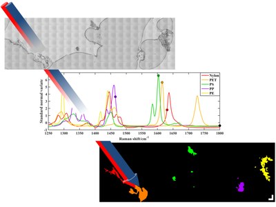
A test sample of microscopic polymer fragments collected on a filter (top). |
The growing amount of plastic micro- and nanoscale particles in canals, lakes, rivers and oceans presents a serious environmental concern. Researchers at Laserlab-Europe partner LaserLaB Amsterdam have built a setup for stimulated Raman scattering microscopy, which can be used to quickly identify the five major types of polymer particles collected on a filter.
In recent years there has been a growing concern regarding environmental microplastics. These polymer particles can have many different chemical compositions, sizes and shapes. They are either added deliberately to cer-tain products such as cosmetics, or can result from plastic waste fragmented by mechanical or photochemical degradation. A large fraction enters our rivers and canals (also because sewage treatment plants do not remove them very efficiently) and ultimately ends up in the marine environment. Large concentrations of floating plastic garbage are known as “plastic soup”, but the much larger numbers of invisible, micro- and nanoscale particles are an even bigger concern. Although the urgency of the problem is clear, methods to detect and identify microplastics are not yet fully developed.
LaserLaB Amsterdam has been involved in several projects related to microplastics and aquatic pollution. Raman spectroscopy is very suitable for the chemical characteri-sation of polymers: when irradiating a sample with a laser and detecting the (very weak) fraction of inelastically scattered light, we obtain a fingerprint spectrum showing the vibrational modes of the molecules in the sample. Different polymer types give very different Raman spectra. The figure shows the Raman spectrum of a white microplastic particle collected from the effluent of a wastewater treat-ment plant. Comparison with reference spectra clearly identifies the polymer as polyethylene.
We are currently looking with a drinking water research facility at the effectiveness of the various purification steps. Raman spectroscopy plays an indispensable role in such projects, but unfortunately the classical mapping approach is quite time-consuming. As a new development, we have therefore built a setup for stimulated Raman scattering (SRS) microscopy, which can speed up the identification of polymer particles collected on a filter by 3-4 orders of magnitude.
For SRS, the sample is irradiated with two (picosecond) laser beams, of which the difference in photon energy matches a vibration of the target molecule. As a result, a signal integration time of the order of microseconds per pixel is sufficient, compared with seconds in the case of conventional Raman spectroscopy. By repeating the SRS image with six different wavenumber settings, the five major polymer types can be distinguished at the fastest mapping speed for environmental samples to date. As a proof-of-principle, we demonstrated this approach for microplastic particles identification in sediment from the Rotterdam harbour area.
Liron Zada & Freek Ariese (LaserLaB Amsterdam)
L. Zada et al., J. Raman Spectroscopy 49: 1136, 2018
Solvation structure and ultrafast motions of hydrated excess protons (MBI, Germany)
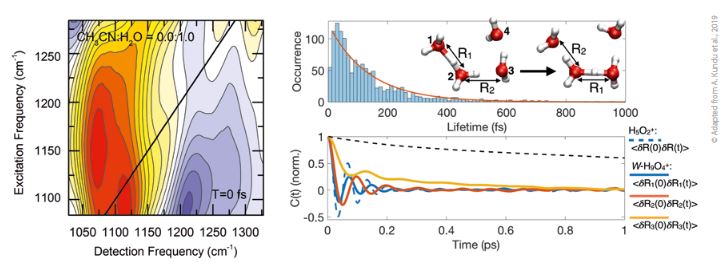 |
| Left: two-dimensional infrared spectrum of excess protons solvated in neat water with ground state bleach. Stimulated emission components shown in red and excited state components shown in blue. Right: timescale of interconversion of the H5O2+ Zundel-motif (marked by R1) within the H3O+ unit obtained from ab-initio molecular dynamics simulations. |
Understanding the solvation shell of hydrated protons is a long-standing open question that is key to proton transport via the Grotthuss mechanism – also known as ‘proton jumping’. Novel experiments at Laserlab-Europe partner MBI shed light on proton solvation structure and dynamics on the ultrafast timescale.
The solvation structure of excess protons in aqueous surrounding is highly relevant to electric properties and for understanding proton transport in liquids and membranes. Despite such overarching importance, the microscopic details of the solvation shell constitute a long-standing unresolved question. Commonly, solvation of an excess proton H+ is discussed within two limiting structures: in the Eigen complex (H9O4+) a central hydronium ion (H3O+) harbours the proton and is surrounded by three water molecules, while in the Zundel cation (H5O2+) the proton is part of a strong hydrogen bond between two flanking water molecules.
In particular the lack of suitable non-invasive experimental observables, as well as structural fluctuations of the solvent molecules on a multitude of timescales, have prevented an unequivocal assignment of the abundance of different solvation structures. By combining nonlinear two-dimensional infrared spectroscopy in the mid-infrared molecular fingerprint region (~ 8 μm) with ab-initio molecular dynamics simulations, we have tracked the genuine proton degrees of freedom in varying acetonitrile/water mixtures, including neat water. The strategy of building the water environment step-by-step provides a benchmark of aqueous solvation geometries via a spectroscopic comparison to excess protons selectively prepared with only a few water molecules in neat acetonitrile. As such, it was demonstrated that for all investigated mixing ratios, excess protons are confined within a low barrier double-minimum potential and are subject to large amplitude displacements due to the fluctuating electric field imposed by the structural fluctuations of the solvent shell. The results thus demonstrate that excess protons in water are predominantly solvated within dimeric water structures, i.e., within a H5O2+ Zundel-motif.
The accompanying simulations reveal how the dimeric water solvation structure interacts with its closest water neighbour in an H7O3+ unit without persistent localiza-tion of the excess proton in a single H3O+ unit. The results suggest that a rearrangement of the hierarchical hydrogen bond structure, accompanied by translocation of the dimeric solvation geometry, is a key element in the microscopic mechanism of proton translocation in aqueous solution. Our investigations thus shed light on the elementary steps of proton translocation in aqueous solution via the Grotthuss mechanism of structural diffusion.
Benjamin P. Fingerhut (MBI)
A. Kundu et al., J. Phys. Chem. Lett. 10: 2287, 2019
Chemistry in ultracold interstellar space: complex behaviour of acids (FELIX, The Netherlands)
 |
|
Two different pathways for the aggregation of HCl(H2O)4 clusters |
Because primordial molecular evolution occurs on icy mantles of dust nanoparticles or on ultracold water clusters in dense interstellar clouds, chemical reactions at ultralow temperatures are of fundamental importance to this process. Using spectroscopic analyses and computer simulations, a team of researchers from Ruhr University Bochum and Laserlab-Europe partner FELIX Laboratory in Nijmegen discovered that at extremely low temperatures proton release from hydrochloric acid (HCl) depends on the order in which water and hydrochloric acid molecules are brought together.
A fundamental question concerning primordial mo-lecular evolution is whether acid/base chemistry, such as discovered in stratospheric clouds, could already be triggered at ultracold ‘stardust conditions’ by dissociation of acids, e.g., HCl, in the absence of thermal and photoin-duced activation.
Solvated acids tend to release a proton, and at room temperature hydrochloric acid (HCl) immediately dissociates when it comes into contact with water molecules. It releases its proton (H+) and one chloride ion (Cl-) remains. However, under ‘stardust conditions’ they display a more complex behaviour. Studying reactions in a stepwise manner in ultracold helium nanodroplets by mass-selective infrared (IR) spectroscopy provides an avenue to mimic these ‘stardust conditions’ in the laboratory.
In a joint experimental and theoretical study, involving researchers from the Cluster of Excellence RESOLVE at Ruhr University Bochum and FELIX Laboratory, H2O molecules have been successively added to HCl, disclosing a unique IR fingerprint at 1337 cm-1 using the FELIX free electron laser. The IR signature signals hydronium (H3O+) formation and, thus, acid dissociation generating solvated protons. In stark contrast, no reaction is observed when reversing the sequence by allowing HCl to interact with preformed small embryonic ice-like clusters.
Ab initio simulations demonstrate that not only reaction stoichiometry but also the reaction sequence needs to be explicitly considered to rationalise ultracold chemistry. One may expect that the results can also be applied to other acids and as such represent a basic principle of chemistry under ultracold conditions.
Britta Redlich (FELIX)
D. Mani et al.: Science Advances 5: eaav8179, 2019
When light meets water: a splash rather than a dip (CLL, Portugal)
 |
|
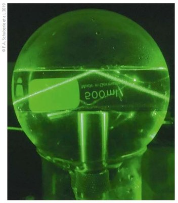
Experimental setup with water and incident light at total |
What happens when light crosses the interface between air and water? Countless experimental attempts to see either a splash or a dip had given contradictory results, but the controversy now seems to have been solved at Laserlab-Europe partner Coimbra Laser Lab (CLL). An experiment involving pulsed light illumination of the air/water interface, under conditions where thermal effects are avoided, clearly shows that the light causes a splash rather than a dip.
Every second, 1035 photons cross the air/water inter-face at the surface of the Earth, change velocity and transfer momentum to that interface. Two competing theories, proposed by Hermann Minkowski and Max Abraham, have been used for over one century to describe what happens when light crosses the air/water interface and slows down. Minkowski proposed a momentum transfer that pushes the water interface up to the air – a splash – whereas Abraham deduced that the momentum transfer pushes the interface in-wards to the water – a dip.
These conflicting theories have motivated many experiments and interpre-tations, but measurements of the momentum trans-ferred at the air/medium in-terface remained elusive. A major experimental difficulty has been the separation of the very minute effect expected for momentum transfer from other possible effects, such as thermal effects due to light absorption. The work at the Coimbra Laser Lab eliminated thermal effects by taking advantage of a remarkable property of water: the thermal expansion coefficient of water is zero at 3.9 °C. Based on three decades of experience in photoa-coustic calorimetry, researchers at CLL used pulsed laser excitation of air/water interfaces at 3.9 °C to measure photoacoustic waves exclusively generated by volume changes at the air/water interface.
The experiments employed the total reflection angle of light to water/air surface, which maximizes the photoacoustic signal generated by photon momentum transfer at the interface. The optical radiation pressure pushes the water/air interface outwards to air and the angular dependence of the photoacoustic waves follows the predictions of Minkowski. There is a splash when the photons hit the water/air interface!
Fabio A Schaberle, Carlos Serpa and Luís G Arnaut (CLL)
F. A. Schaberle et al., New Journal of Physics 21: 033013, 2019.
Monitoring water quality using endogenous fluorescence of algae (ILC, Slovakia)
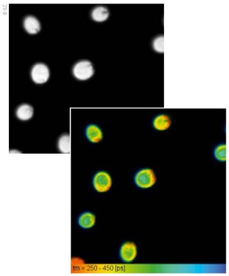 |
| Fluorescence lifetime imaging (FLIM) of endogenous fluorescence in marine algae Dunaliella, following excitation by a 475 nm picosecond laser, recorded by a TCSPC system. Intensity im-age (left); lifetime distribution image (right), shown in the range 250-450 ps, demonstrates the presence of at least 2 fluorescence lifetimes in control conditions. |
Photosynthetic pigments in algae are sensitive to changes in their environment, thus acting as natural biosensors that reflect changes in water quality. At Laserlab-Europe partner International Laser Center (ILC) confocal microscopy and time-resolved fluorescence imaging are employed to study the reaction of algae to various kinds of stress. The research should lead to new methods for monitoring water quality.
Acidification of the aquatic environment presents an important environmental stress factor, as under the influence of industrial pollution acid rain can reach a pH of below 5, severely impacting mainly coastal water. Pollution by heavy metals constitutes a hazard to aquatic organisms as well.
Algae play a crucial role in aquatic ecology, producing oxygen and serving as the food base for almost all aquatic life. We evaluate responses of the endogenous fluorescence related to chlorophylls, as well as other pigments, such as flavonoids and carotenoids, in algae. Pigments of various algae, namely seawater algae Dunaliellaand fresh water algae Chlorella sp., as well as phototropic Euglenas are studied by advanced spectroscopy and microscopy methods in their natural environments, but also in response to environmental changes, namely pH modification, or presence of heavy metals – more specifically cadmium.
Employing advanced spectrally-resolved confocal microscopy and time-resolved fluorescence imaging, we study the fast (in terms of minutes), as well as the long-term (in terms of weeks) responsiveness under stress conditions of the algae, but this approach can also be used to monitor changes in water moss, or higher plants. Deep understanding of algal cell response to stressors will be helpful for monitoring water quality, and can become essential in biosensing and thus predicting how ecosystems may be affected by water pollution, climate change and other human activities.
In more detail, fluorescence lifetime (FLIM) autofluorescence images are recorded using the time-correlated sin-gle photon counting (TCSPC) technique following excitation with a 475 nm picosecond laser diode. The laser beam is reflected to the sample through an epifluorescence path of a laser scanning confocal microscope. The emitted fluorescence is separated from laser excitation using a long pass filter 500 nm and detected by a photomultiplier array. This work is done in the common lab of biophotonic technologies of the International Laser Center and the University of Ss. Cyril and Methodius in Trnava, Slovakia, in collaboration with Ruder Boskovic Institute in Zagreb, Croatia.
Alzbeta Marcek Chorvatova (ILC and University of Ss. Cyril and Methodius)
N. Ivosevic DeNardis et. al., European Biophysics Journal 48: 231, 2019
Two types of water revealed by ultrafast spectroscopy (LENS, Italy)
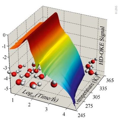 |
| Heterodyne Detected – Optical Kerr Effect data of liquid water at different temperatures |
Water might actually come in two distinct forms, which could explain its peculiar behaviour compared with other liquids. Experiments using ultrafast laser spectroscopy, performed at Laserlab-Europe partner LENS, provide evidence of the coexistence of two local structural configurations of water molecules, which supports the hypothesis of high-density and low-density forms of water.
Water can remain liquid below its freezing point, en-tering into a metastable phase known as “supercooled water”. In this water phase, a series of physical characteristics show anomalous behaviours that distinguish it from other liquids (e.g. the density has a maximum at 4°C). Despite the relative simplicity of water molecules, its liquid phase remains an open problem for liquid physics; the strong net-work of hydrogen bonds formed by the molecules support extended and collective interactions, which are very hard to describe by physical models.
One of the most credited models assumes that water is actually made up of two different types of liquid: low-density water and high-density water. The former is characterized by tetrahedral local intermolecular structures, very similar to those found in ice, and the latter by less ordered and more compact structures. According to this model, at low temperatures these two water forms are distinct and separated by a phase transition, while at higher temperatures they interpenetrate and continually exchange in extremely rapid timeframes, typically 10-12 seconds.
Experimental detection of these two water forms is made particularly difficult precisely by the speed and frequency of the rearrangements of the local liquid structures. To date there is still no definitive proof of the existence of these two water forms. However, in an ongoing campaign, a group of researchers at the European Labo-ratory for Non-Linear Spectroscopy (LENS, University of Florence) is undertaking a series of experimental studies using ultrafast spectroscopy techniques based on femto-second laser sources.
Measuring the dynamics of liquid water at temperatures down to -28 °C, these studies show that vibrational and structural dynamics of the water molecules reveal the presence of two major molecular organizations: one characterized by a high order of tetrahedral hydrogen bonds, while the other presents strong local distortions of the lattice. These two types of local organization of water molecules can be interpreted as evidence of the existence of water of low and high density.
Renato Torre (LENS)
A. Taschin et al., submitted, 2019A.
Taschin et al., Nat. Commun. 4: 2401, 2013
Micro-LIBS imaging of mollusc shells: a window to the past (IESL-FORTH)
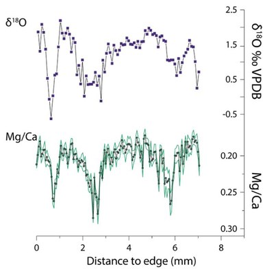 |
| Records showing the good correlation of expensive oxygen isotope records (above) and rapid LIBS records. |
In a recent article in Scientific Reports[1], resulting from a Laserlab-Europe transnational access project, scientists at IESL-FORTH, collaborating with the Max Planck Institute for the Science of Human History and the School of Geography, University of Melbourne, have shown that LIBS microscopy enables extensive elemental mapping of mollusc shells, providing an insight into the rich environmental archives contained in such specimens. This is a major step forward for the exploitation of mollusc archives to study climate in a range of applications, including ocean circulation, glacial/interglacial climates, and anthropogenic climate change.
Shellfish have played a significant role in the diet of pre-historic coastal populations, providing valuable nutrients. They are a common find at archaeological sites all over the world, usually in huge numbers, and researchers have long explored how they could be used to make inferences about the environments that humans experienced at those locations in the past. However, although techniques have been developed to extract valuable climate-related information from shells, it has been too expensive to obtain data on a scale beyond individual and isolated records. The current study presents a technique to use rapid laser imaging to increase the number of analysed shell records to previously unknown scales, and thereby greatly expand the time periods and accuracy of the reconstructed records.
The effort was led by Niklas Hausmann at IESL-FORTH in the framework of his Marie Skłodowska Curie fellowship project ACCELERATE (www.accelerate-project.com). He joined the research group of Demetrios Anglos (University of Crete and IESL-FORTH) and collaborated with Panagiotis Siozos, senior researcher, and Andreas Lemonis, automation and programming expert, developing an automated scanning micro-LIBS (laser-induced breakdown spectros-copy) apparatus that enabled mapping of large numbers of shells of the course of the project.
It is important to note that this research emerged from a Laserlab-Europe access project. It was in December 2013 that Hausmann, then a PhD student at the University of York, UK, approached Costas Fotakis and Demetrios Anglos at the Ultraviolet Laser Facility-FORTH inquiring on the possibility to use their laser for carrying out oxygen isotope analysis on shells. After an access proposal was approved, Niklas Hausmann visited ULF-FORTH and carried out a series of exploratory experiments in April-May 2014.
Following two weeks of hard work, Hausmann and his co-workers were able to obtain the first LIBS images showing variations of the magnesium to calcium (Mg/Ca) concentration on sections of mollusc shells[2, 3]. These variations, traced along the shell growth lines, were consistent with seasonal temperature variations, showing the potential of micro-LIBS as a new tool for obtaining high-resolution elemental images of shell surfaces.
This success was enough to spark a more consistent effort to investigate LIBS mapping in detail, and led to the ACCELERATE project. Within this project, mollusc shells from a range of countries and archaeological sites were analysed, covering 20 countries on all continents and ranging back into past time periods with shells collected by Neanderthals. Most shells, however, dated to modern periods, where the elemental composition could be compared to readings from instrumental data, as was the case for the study in Scientific Reports[1]. Here LIBS allowed to analyse quickly and at low cost shells from across the Mediterranean in a wide and multi-regional study that compared different localities, rather than only one, resolving major concerns in the field regarding the improvement of rapid and low-cost climate proxies such as Mg/Ca ratios in shells.
So far, it had been hard to link such elemental ratios of shell carbonate to sea surface temperatures because the interpretation of the measurements was hampered by variations in study location as well as vital or physiologi-cal effects between and within individual mollusc specimens. Having identified the Mediterranean limpet Patella caerulea as an ideal species to assess elemental variations through LIBS, elemental maps and line scans of 19 shell specimens from 9 sites were produced for the study.
The Mg/Ca ratio maps of almost all specimens show repeating bands of high and low intensity throughout the entire length of growth increments, corresponding to the seasonal changes in sea surface temperature known from satellite measurements. Only two shells – the ones that had grown for less than a year – lacked these bands. As many as 8 out of the 19 specimens, however, showed ad-ditional Mg/Ca patterns unrelated to sea surface temperature, and in two of those specimens this prevented reliable temperature reconstruction. Such problematic specimens would skew the sea surface temperature record if measured without being recognised. The observed seasonal changes in Mg/Ca ratios obtained by LIBS allow archaeologists to determine at relatively low costs in which season the specimen has been consumed, which is of importance for the study of past human and animal behaviour.
Hausmann is now seeking funding to set up a dedicated micro-LIBS facility for large-scale mollusc screening, based on the outcomes of the ACCELERATE project, which did not only find new ways of analysing temperature changes, but also to reveal growth structures within shells. These can be key to efficiently plan analyses using other geochemical proxies, such as oxygen isotope analysis or clumped isotope analysis, which are more expensive.
In addition to molluscs, there are other carbonate archives with their own interesting research trajectories that LIBS mapping can address. Stalagmite and corals have previously been shown to provide important elemental records of climate change and would benefit from rapid analysis or screening prior to more detailed geochemical studies. The team at FORTH investigates these potential applications.
Niklas Hausman (University of York),Panagiotis Siozos & Demetrios Anglos(IESL-FORTH)
1. N. Hausmann et al., Scientific Reports 9: 3698, 2019
2. N. Hausmann et al., J. Anal. At. Spectrom. 32: 1467, 2017
3. N. Hausmann et al., pp 725-740, in: N. Rasul, I. Stewart (eds), Geological Setting, Palaeoenvironment and Archaeology of the Red Sea, Springer, Cham, 2019